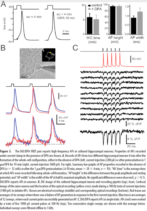
I have been itching to post about this work since David DiGregorio presented it at a meeting at Janelia last year. His group’s results, Submillisecond Optical Reporting of Membrane Potentials In Situ Using a Neuronal Trace Dye, were published in the Journal of Neuroscience last week. Their method of optical voltage sensing is the first one that looks like its ready for “prime-time” action outside of the labs of developers of these sorts of techniques. It has sufficient speed (<1 ms resolution), sensitivity (25% dF/F per 100mV), and limited membrane perturbation to see single action potentials, without dramatically altering the shape of these currents.

Membrane depolarization causes DPA to rapidly partition to the inner membrane leaflet, quenching DiO.
Like previous methods, Bradley et al. use voltage-dependent membrane partitioning of dipicrylamine (DPA), a charged small molecule, to quench a fluorophore via FRET. Previously, high-concentrations of DPA were required to have a reasonable signal change, which caused toxicity, increased membrane capacitance and slowed voltage transients. By using DiO, a lipophilic neuronal tracer, as the fluorophore, the DPA concentration could be reduced to 1uM, while retaining sufficient optical sensitivity for action potential detection.

I’m interested in, but unfamiliar with the new optical imaging breakthroughs. Is this designed for single-cell recording in vitro, or anesthetized in vivo, or could I stick a CCD chip in the middle of a rat brain and pick up multiple unit activities?
Probably limited to cultures and slice at this point since you need controlled delivery of the small molecules. There are some genetically encoded versions that will potentially be useful in vivo, but don’t have the same signal/noise or kinetics.
This is a FRET based approach. The optical traces of action potential remain noisy even after signal average. There is still space for improvement.
Personally I perfer use the fast voltage-sensitive dyes. ANNINE-6plus would be my first choice. It has fast response (could be nanosecond, often limited by your microscope or other equipment) and high voltage sensitivity (>30% per 100 mV membrane potential change). More importantly, ANNINE-6plus is water solable. It is very easy to peropare the dye solution and to stain the preparation.
I cannot comment on its in vivo application. Most likely, it will work. It works perfectly on different preparations, cultured cells, slice, or whole organ.
As to single cell or multiple-site recording, you can use either spin-disc microscope (or similar systems) or optical mapping systems with CCD camera or PDA (photo diode array). The optical mapping systems have been used in many studies of heart research.
I am more than happy to share with you my experience on voltage sensitive dyes and their applications in heart research and neuroscience.
The work by DiGregorio used FRET and required two different compounds. Compared to fast potentiometric dyes such as ANNINE-6plus, it should be much harder to deliver these molecules to the targeted tissue. It is easy to stain the tissue with ANNINE-6plus, using ~10 minutes incubation for cultures or ~10 minutes perfusion for tissue. I don’t have experience with brain/ganglia slices, but I worked with intact dorsal ganglia and mouse heart before. Staining these specimens with ANNINE-6plus is pretty easy and the quality is usually very good.
ANNINE-6plus also has fast response, in ns range, which should be faster than FRET. However, the temporal resolution is often limited by the imaging instruments rather than the dye molecules. With regard to voltage sensitivity, ANNINE-6plus is definitely the dye of choice with the highest voltage sensitive to date.
By the way, the APs in the figures are results from signal average rather than single-sweep traces. You need to have some signal processing algorithms to handle this specific narrow band signal with low signal-to-noise ratio. I designed an algorithm which works pretty well on this type of signal.
Hi!!
I’m trying to find a dye for my first experiment with neurons. But, where can I order Annine-6plus???
Thanks a lot.
Hi, great blog!
@ Daniela, Hi,
To my knowledge, ANNINE-6plus is not commercially available, otherwise I’m also interested in purchasing it.
Cheers!
ANNINE-6plus has been commercially available since 2008.