Journal Club – In Vivo Inhibition Dynamics

18 02 2010

Inhibition has a powerful role shaping the network dynamics of the cortex, but most studies of inhibitory circuitry are done in brain slice or anesthetized animals. In Membrane potential dynamics of GABAergic neurons in barrel cortex of behaving mice, Gentet et al use two-photon imaging to guide dual, whole-cell patch clamp of inhibitory and excitatory neurons in the mouse barrel cortex.  These mice are head fixed, but awake and naturally whisking.  The authors can then see how the membrane dynamics of both subthreshold and suprathreshold voltages are correlated across pairs of cells.  Differences between the correlations for excitatory and inhibitory neurons shed light on how cortical circuitry processes sensory information in natural brain states.

For Journal Club #5, Mac Hooks, a post-doc here at Janelia working with Gordon Shepard and Karel Svoboda, walks us through these results.  Also, there is a video introduction of the work by the lab head of the paper, Carl Petersen, provided by Cell Press.


Gentet, L., Avermann, M., Matyas, F., Staiger, J., & Petersen, C. (2010). Membrane Potential Dynamics of GABAergic Neurons in the Barrel Cortex of Behaving Mice Neuron, 65 (3), 422-435 DOI: 10.1016/j.neuron.2010.01.006


Playing Quake with a Real Mouse

14 10 2009

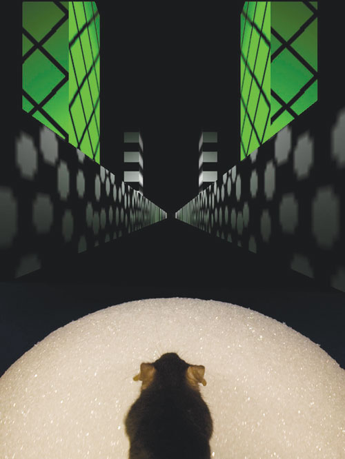
Most people play Quake with a computer mouse, but researchers in David Tank’s lab at Princeton have done it with a living mouse, AND they are recording the intracellular activity of individual neurons of the mouse during the gaming session. As reported in Intracellular dynamics of hippocampal place cells during virtual navigation, the virtual reality environment of the video game was sufficiently realistic to generate place cell activity in the mouse’s hippocampus. 

Now where did I see that cheese power-up?

Now where did I see that cheese power-up?

Place cells modulate their activity dependent on the location the mouse is at. They have mostly been identified with extracellular recordings in freely moving mice. Extracellular recording only permits the detection of the rates of action potential firing, rather then the subtle intracellular voltage changes that could help explain the mechanism of place cell activity generation.  A few pioneers, such as Albert “my greatest strength is a tremendous capacity for boredom” Lee, have recorded intracellularly in freely moving animals, but these experiments are fiendishly difficult, as the motion of the animal’s head tends to break the seal on the recorded neuron.  Only a few cells have been recorded in that manner for more than a few minutes, though the success rate has been improving recently.  

Experimental setup

Experimental setup

In Chris Harvey’s technique, they fix the head of the mouse to a bar and let the mouse walk on a floating ball, while a virtual reality screen is projected in the mouse’s field of view.  The motion of the ball controls the motion on the screen.  The head never moves, so intracellular recordings can be made relatively easily and held for long periods of time.

The authors find three characteristics of place cell activity that could explain their generation and function.

“An asymmetric ramp-like depolarization of the baseline membrane potential, an increase in the amplitude of intracellular theta oscillations, and a phase precession of the intracellular theta oscillation relative to the extracellularly recorded theta rhythm.” 

Intracellular voltage dynamics in place cells

Intracellular voltage dynamics in place cells

These could be used to explain how place cells remap their selectivity when a mouse (or a human) moves into a new environment.  This also could be used to do more in depth studies of the mental replay of place locations that has been previously recorded in the activity patterns of the hippocampus.  The technique itself is about as sexy as neuroscience gets.  Unfortunately, this paper also provides an additional piece of evidence for Karel to use in motivating lab post-docs, “Look at Chris, he left the lab after you got here and already has a Nature article…”‘


Voltage imaging with sub-millisecond, single-action potential resolution

27 07 2009

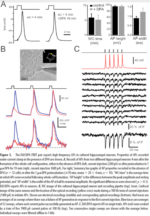
I have been itching to post about this work since David DiGregorio presented it at a meeting at Janelia last year. His group’s results, Submillisecond Optical Reporting of Membrane Potentials In Situ Using a Neuronal Trace Dye, were published in the Journal of Neuroscience last week.  Their method of optical voltage sensing is the first one that looks like its ready for “prime-time” action outside of the labs of developers of these sorts of techniques.  It has sufficient speed (<1 ms resolution), sensitivity (25% dF/F per 100mV), and limited membrane perturbation to see single action potentials, without dramatically altering the shape of these currents.  


Membrane depolarization causes DPA to rapidly partition to the inner membrane leaflet, quenching DiO.

Membrane depolarization causes DPA to rapidly partition to the inner membrane leaflet, quenching DiO.


Like previous methods, Bradley et al. use voltage-dependent membrane partitioning of dipicrylamine (DPA), a charged small molecule, to quench a fluorophore via FRET.  Previously, high-concentrations of DPA were required to have a reasonable signal change, which caused toxicity, increased membrane capacitance and slowed voltage transients.  By using DiO, a lipophilic neuronal tracer, as the fluorophore, the DPA concentration could be reduced to 1uM, while retaining sufficient optical sensitivity for action potential detection.

Picture 9


Sensing salty currents with Mermaids

16 07 2008

A new genetically-encoded voltage sensor paper is out from a friend and former mentor of mine, Atsushi Miyawaki. One memorable moment when working in his lab during the RIKEN summer program of 2002 was when Atsushi took me into his office and whipped out a custom green laser pointer. These had been banned in Japan, as fans would shine their powerful light into the eyes of pitchers and batters at baseball games. Atsushi was really proud of his. He smiled and then started sweeping the light point over the rocks in his fishtank. Each ‘rock’ was actually coral his lab had collected from fluorescent protein hunting trips, and each glowed a different color when the green light hit it. He has been putting these novel discoveries to good use.

In Improving membrane voltage measurements using FRET with new fluorescent proteins, Tsutsui et. al take two fluorescent proteins discovered and engineered by the Miyawaki lab, mUKG and mKOk, and graft them onto the Ci-VSP scaffold used in VSFP2.1 (also developed at RIKEN).  The green and orange fluorescent proteins undergo significant FRET transfer which is voltage dependent.  They get 40% dR/R per 100mV with a 2 component association rate of around 10 and 200ms. Unsurprisingly, the kinetics speed up at physiological temperatures to 5-20ms on and off.  They are able to pick up single pseudo-action potentials in Neuro2A cells, though the response is highly filtered. They are also able to see very clear spontaneous waves of potential change in cardiomyocytes (23% dR/R) and single spikes in cultured neurons (2% dR/R for 1AP). They dub this voltage sensor “Mermaid”.

The authors state that they used the new FPs due to their improved photostability and especially pH resistance. 

Additionally, because Aequorea GFP variants are pH-sensitive, and neuronal activity causes considerable acidification, the responses of sensors to depolarization in intact neurons may be overwhelmed by sustained changes resulting from acidification.

Granted that mOrange2 is pretty pH-sensitive, but I’m not sure this is a real issue, or a potential issue to justify using their new FPs.  From the spectra of mUKG vs. EGFP, it would seem that EGFP’s 10nm further redshifted emission would be a superior FRET pair for mKOk.  It smells like there may be a bit of bundling of various independent projects into this paper.  However, they do make a good point that this pair will have a different preferred dipole orientation than existing FRET pairs, which could lead to improved performance in some constructs.  

Things I’m still wondering :

  • Have they tried using the improved VSFP3.1 scaffold? This was shown to be much faster than 2.1.  I suspect the mUKG is not as tolerant to C-terminal truncation than CFP and GFP.  
  • What about using EGFP as the donor?  Could you then use the VSFP3.1 scaffold?
  • Is there a rapid non-FRET quenching of the donor upon depolarization as seen in VSFP3.1?
  • Why is the single wavelength fluorescence increasing in both channels in figure 2d?  Is there some photoactivation going on?
  • I’d love to see a head to head comparison of VSFP3.1 and Mermaid under identical conditions. Also responses in brain slice at physiological temperatures.


Voltage sensitive imaging powering up

8 07 2008

I’m starting to come around on voltage imaging. I haven’t been a fan of it for a number of reasons.

  • The response sizes suck.  Classic dyes and genetically encoded systems get a few percent fluorescence change at best. 
  • The response speeds suck. Measuring continuous current injections from -100mV to +150mV is not very interesting.  Action potentials are interesting.  But they are fast.
  • Toxicity. The dyes kill neurons, or strongly perturb their electrical properties.

OK, voltage-sensitive imaging isn’t totally useless, for example see Carl Petersen’s recent paper on Spatiotemporal Dynamics of Cortical Sensorimotor Integration in Behaving Mice (2007). But if the above problems could be solved, then voltage sensitive imaging would be a strong competitor to calcium imaging for the non-invasive, high-resolution monitoring of patterns of network activity. There has been considerable progress ameliorating these problems in the past few years, much of it by a consortium of labs (Isacoff, Knöpfel, Bezanilla, Miesenböck, and others) focused on these issues. (Umlaut’s apparently help in this field).

First, let’s look at a minor breakthrough for the fully genetically-encoded strategy.  In Engineering and Characterization of an Enhanced Fluorescent Protein Voltag Sensor (2007), The Knöpfel group tagged the recently discovered voltage sensitive phosphotase (Ci-VSP) with CFP and YFP FRET pairs in place of the phosphotase domain. This tagged protein expressed at the membrane much more efficiently than previous genetically encoded voltage sensors based on potassium channel subunits. By injecting physiological voltage changes and averaging 50-90 traces, they were able to pull out a few percent ratio change from a brief series of action potentials. Single spikes were resolvable.  Although this sensor (VSFP2.1) was pretty slow (tau > 10ms), this new substrate looked promising for future sensor development.

They have since sped the response up.  In Engineering of a Genetically Encodable Fluorescent Voltage Sensor Exploiting Fast Ci-VSP Voltage-Sensing Movements (2008 ), they determined that the gating motion of the voltage sensing component was very fast (~1ms), while the fluorescence change was slow (~100ms). So they did what any good FRET tinkerer would do, chop away at the linkers between FP components.  The sensor response improved, and they noticed that there was a disconnect between the speed of the CFP and YFP responses. Not only was CFP decreasing from enhanced FRET, it was being directly quenched by interactions with the lipid membrane. Chopping off the YFP from the the construct then dramatically increased the speed of the CFP quench. This improved sensor, VSFP3.1 has an activation time constant of 1.3ms, though it’s response magnitude is still quite small (a few % dF/F).

A hybrid approach to measuring electrical activity in genetically specified neurons (2005) has a much greater response magnitude. Pancho Bezanilla’s group exploited the rapid, voltage-dependent translocation of the small molecule quencher dipicrylamine (DPA) through the plasma membrane to change the fluorescence of membrane-teathered GFP in a voltage-dependent manner. Responses of the hybrid voltage sensor (hVOS) were relatively large (34% per 100mV) and fast (0.5ms). Single action potentials were detectable without averaging.  However, since DPA is a charged molecule, it significantly increased the capacitance of the membrane. The levels of DPA required to see large responses inhibited action potentials and were intolerable to neurons.

Last month in Rational Optimization and Imaging In Vivo of a Genetically Encoded Optical Voltage Reporter (2008 ), Sjulson and Miesenböck reported optimized parameters for the hVOS approach. They built a quantitative model of the quenching effects of DPA on membrane-teathered GFP.  The quenching is limited by the distance the DPA can approach the chromophore of GFP.  Only the closest DPA molecule to the chromophore significantly contributes to a GFP’s quenching. After lots of pretty heat maps and graphs, the model tells them to chop off the tail of EGFP to bring the C-terminal tethering sequence closer to chromophore. I should note that an 11 amino acid C-terminal truncation of ECFP has improved the response of a tremendous number of FRET reporters and has been standard practice for the last 8 years. By shortening the linker they manage to triple the response size. I’d suggest, if they haven’t already, to lop off another six amino acids (end the EGFP with …LEFVTAA) and see if works.  EGFP and ECFP usually tolerate it.

Using this optimized reporter, they are able to reduce DPA concentrations to levels that are usable in vivo, at least for a few minutes. They record fast optical responses to electrical activity in the Drosophila antennal lobe using 2uM DPA.  But after a few minutes, the DPA loaded neurons become strongly inhibited.


The bottom line? Voltage-sensitive imaging has seen big progress in the last few years, but still has a long way to go to gently record single APs in a dish or in vivo. Or does it?  I’m hearing whispers that a different group has developed a synthetic dye technique that is getting >10% dF/F to single APs with millisecond response times. Is it the real deal? Watch this space…


Blog Roundup

27 02 2008

Here’s a quick overview of some posts that got my attention in the last month…

Neurodudes has a brief writeup of video-rate superresolution imaging from Stefan Hell’s group. I don’t have access to Science Express PDF’s through our institutional subscription (how much must they be charging for that?), making a full writeup impossible. But you can at least check out the abstract and supporting info here. Video-Rate Far-Field Optical Nanoscopy Dissects Synaptic Vesicle Movement (Westphal et al.). The optical resolution isn’t quite as good as PALM or STORM, but the speed of acquisition is fantastic, permitting its use on dynamic living processes.

Eric Thomson from Neurochannels has posted a detailed Journal Club style review at Nature Network of the paper Spatiotemporal Dynamics of Cortical Sensorimotor Integration in Behaving Mice (Ferezou et al.) from Carl Petersen’s group. Using voltage-sensitive dyes they show the timing and spreading of activity from the sensory to motor cortex, following whisker stimulation in awake mice.

Biosingularity reports on results from Susumu Tonegawa’s group published in Science Express as Transgenic Inhibition of Synaptic Transmission Reveals Role of CA3 Output in Hippocampal Learning (Nakashiba et al.). They use a novel method, doxycycline-inhibited circuit exocytosis-knockdown (DICE-K), to transiently and selectively shut down the tri-synaptic pathway of the hippocampus (ER->DG->CA3->CA1->ER), while leaving the monosynaptic pathway (ER->CA1->ER) intact. These mice can still learn incrementally, but one-trial contextual learning and pattern completion recall is wiped out.