G-CaMP5 is finally published!

3 10 2012

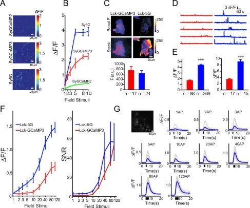
The paper on G-CaMP5 has been published.

Optimization of a GCaMP Calcium Indicator for Neural Activity Imaging

Genetically encoded calcium indicators (GECIs) are powerful tools for systems neuroscience. Recent efforts in protein engineering have significantly increased the performance of GECIs. The state-of-the art single-wavelength GECI, GCaMP3, has been deployed in a number of model organisms and can reliably detect three or more action potentials in short bursts in several systems in vivo. Through protein structure determination, targeted mutagenesis, high-throughput screening, and a battery of in vitro assays, we have increased the dynamic range of GCaMP3 by severalfold, creating a family of “GCaMP5” sensors. We tested GCaMP5s in several systems: cultured neurons and astrocytes, mouse retina, and in vivo in Caenorhabditischemosensory neurons, Drosophila larval neuromuscular junction and adult antennal lobe, zebrafish retina and tectum, and mouse visual cortex. Signal-to-noise ratio was improved by at least 2- to 3-fold. In the visual cortex, two GCaMP5 variants detected twice as many visual stimulus-responsive cells as GCaMP3. By combining in vivoimaging with electrophysiology we show that GCaMP5 fluorescence provides a more reliable measure of neuronal activity than its predecessor GCaMP3. GCaMP5 allows more sensitive detection of neural activity in vivo and may find widespread applications for cellular imaging in general.

Image

This is the best fully-characterized GECI available, but publication of the paper was repeatedly delayed. Why? Because reviewers viewed it as ‘too incremental’ of an upgrade, and not worth publishing in a prominent journal (no, I’m not talking Nature or Science level) when the plasmids are already available.

A friendly suggestion for authors and future GCaMP6+ reviewers : You can’t have it both ways. If you want access to the best molecular tools before publication (GCaMP5 has been available for over a YEAR), you cannot turn around and say its not worth publishing because you already have the plasmid. Multiple post-docs spent years of their lives developing and carefully testing this tool. They deserve a quality publication for their efforts. Furthermore, the rigorous performance data collected NEEDS to be available to current and future users. Finally, there is no doubt that this will be a highly cited and viewed paper in whatever journal were to publish it. Our GCaMP3 paper already has 182 citations in less than 3 years, this may do even better.


Rapid warping of two-photon illumination wavefronts

16 02 2011

A short paper in Optics Express looks interesting.  In A high speed wavefront determination method based on spatial frequency modulations for focusing light through random scattering media, Meng Cui presents a method for rapidly determining the optimal wavefront to ‘cancel out’ the scattering when 785nm light passes through turbid media.  In his example, a glass diffuser was used, but the clear goal for this work is to replace the glass with a brain.

To understand why this is so important for in vivo two-photon imaging, let’s review how 2-p imaging works. Light from a laser is focused to a point and swept across the field in a raster. The resulting fluorescence is of a different wavelength and can thus be filtered out from the excitation light. For each voxel, all the fluorescence that re-enters the objective is collected, regardless of its source.  The total amount of fluorescence collected for that timepoint in the sweep is assigned as the brightness of that voxel. Since the user knows where the laser was being aimed, scattering of fluorescence emission may reduce the brightness but will not blur the image.  However, scattering of the excitation light can dramatically reduce the excitation at the target voxel while increasing the off-target excitation of its neighbors. This causes a rapid increase in background fluorescence and blur at increasing brain depth.

The vasculature was labeled by injecting flourescein dextran into the circulatory stream. The light source was a regenerative amplifier. ‘‘0 mm’’ corresponds to the top of the brain. Left, XZ projection. Right, examples of XY projections. Note the increase in background fluo- rescence deeper than 600 mm in the brain due to out-of-focus 2PE. (Theer et al., 2003)

Previous reports work has shown that one can use adaptive optics to adjust the phase of the wavefront of the excitation light to correct for the scattering of the excitation.  However, determination of the optimal wavefront for a field of view took minutes, which could be problematic for imaging in an awake animal.  Any changes in the precise position of the brain might change the optimal wavefront.  Ideally, one would want a system that could optimize the wavefront every second, or even before every frame of acquisition (typically 4-8 Hz in a raster scan in vivo experiment)

Scattering in the brain warps two-photon excitation light, but adaptive optics can correct this.

I’ll let Meng Cui explain the technique in his own words

Elastic scattering is the dominant factor limiting the optical imaging depth in tissues. Take gray matter as an example, at 800 nm the scattering coefficient is 77 /cm and the absorption coefficient is 0.2 / cm. If there is a way to suppress scattering, the optical imaging depth could be greatly improved. Despite the apparent randomness, scattering is a deterministic process. A properly engineered wave can propagate inside scattering media and form a focus, a well understood phenomenon in the time reversal and optical phase conjugation (OPC) studies…

For applications on biological tissues, acquisition time on the order of one millisecond (ms) per degree of freedom is desired. Deformable mirrors can provide a high modulation speed. However the degrees of freedom are rather limited. A phase-only SLM can provide about one million degrees of freedom at a much lower modulation speed. In this work, I present a novel method, capable of providing as many degrees of freedom as a SLM with a data acquisition time of one ms per degree of freedom. The method was employed to focus light through a random scattering medium with a 400 ms total data acquisition time, ~three orders of magnitude faster than the previous report [25].

The essence of a COAT system is to phase modulate different input spatial modes while detecting the output signal from the target. To greatly improve the operation speed, the experiment requires a device that can provide fast phase modulation and can access a large number of spatial modes very quickly. To meet these two requirements, a pair of scanning Galvanometer mirrors was used to quickly visit different modes in the spatial frequency domain or k space, and a frequency shifted reference beam was provided for a heterodyne detection. The wavefront profile was first determined in k space and then transformed to the spatial domain. The spatial phase profile was displayed on a SLM to focus light onto the target. In such a design, the number of degrees of freedom is limited by the number of pixels on the SLM and the experiment speed is determined by the scanning mirror speed…

Compared to existing techniques, the reported method can provide both a high operation speed and a large number of degrees of freedom. In the current design, the operation speed is limited by the scanning mirror speed and the maximum number of degrees of freedom is limited by the SLM pixel number. In this demonstration, 400 spatial modes in k space were visited and the determined phase profile was displayed on the SLM. Depending on the scattering property of the media, more (up to 1920 x 1080) or less number of degrees of freedom can be used to optimize the focus quality and the operation speed.

Using a stepwise position scanning, the method achieves an operation speed of one ms (400 μs transition time + 600 μs recording time) per spatial mode, ~three orders of magnitude faster than the previous report. Using a continuous position scanning and a faster position scanner such as resonant scanning mirrors, polygon mirror scanners, or acousto-optic deflectors, the operation speed can be potentially increased by at least one order of magnitude. It is anticipated that the reported technique will find a broad range of applications in biomedical deep tissue imaging.


Backyard Brains Homebrew Ephys Rigs

6 08 2010

News of a cool new toy comes from a colleague’s recent trip out to the MBL @ Woods Hole.  It is the perfect gift to spark the curiosity of a budding young (or old) neuroscientist. Backyard Brains makes the world’s best value electrophysiology rigs. The SpikerBox comes pre-assembled for $100 or build your own from a bunch of parts for $50.

These rigs are surprisingly powerful. You can go out in the yard, catch a bug (or buy a cockroach), strap one on and start recording neuromuscular potentials. The box interfaces with gorgeous iPhone/iPad so you can hear, see and record the action potentials. They don’t make a big point of this, but you can also wire it up so that the piezo-speaker can drive an electrical stimulator of the cockroach’s leg. You can make the leg twitch to whatever signal input you give it. The educational possibilities of this gear is really limitless.


Cell Cycle Visualization in Development

13 03 2010

Atsushi Miyawaki’s lab has developed a series of neat tools for visualizing cell cycle progress.

For zebrafish, the zFucci system consists of two fluorescent proteins, mKO2 and mAG, that are fused to Cdt1 and geminin genes.  Cell cycle- regulated proteolysis of these fusion proteins causes each cell to display orange fluorescence in G1 phase nuclei and green fluorescence in both the nucleus and cytoplasm of S/G2/M phase cells.

Video of cell cycle transitions in culture. Click for the video.

The last time I saw Atsushi give a talk, he showed an incredible time lapse video from the zebrafish cleavage stage that I haven’t been able to find online.  However, here is a video from later in development of the zebrafish that is still pretty remarkable.

Development of a zebrafish visualized by zFucci. Click to see the video.

This two component system has been adapted for watching the transition from neural stem cells to differentiated neurons in living mice. The Color-Timer system uses double transgenics with the fluorescent protein KOr fused to nestin and EGFP fused to doublecortin.  In this system, neural stem cells fluoresce orange, while newly differentiated neurons fluoresce green.

The cerebral cortex of an E14.5 double Tg mouse embryo of nestin/KOr was time-lapse imaged. Click for video

Sugiyama, M., Sakaue-Sawano, A., Iimura, T., Fukami, K., Kitaguchi, T., Kawakami, K., Okamoto, H., Higashijima, S., & Miyawaki, A. (2009). Illuminating cell-cycle progression in the developing zebrafish embryo Proceedings of the National Academy of Sciences, 106 (49), 20812-20817 DOI: 10.1073/pnas.0906464106

Kanki, H., Shimabukuro, M., Miyawaki, A., & Okano, H. (2010). “Color Timer” mice: visualization of neuronal differentiation with fluorescent proteins Molecular Brain, 3 (1) DOI: 10.1186/1756-6606-3-5


Journal Scan – Calcium Imaging in Auditory and Visual Cortex

4 03 2010

A few papers on in vivo calcium imaging have just come out and are worth a careful read.

The first two examine the fine organization of layer 2/3 of the mouse auditory cortex.  The canonical view of auditory cortex organization is that neurons are arranged in a tonotopic pattern, with a smooth gradient in auditory frequency tuning across the surface of the cortex.  Using two-photon imaging in anesthetized mice, the groups saw that, while there was an overall gradient, the tuning of neighboring neurons was highly variable.  These are similar results to what Sato et al and Kerr et al found in the whisker barrel cortex back in 2007.  Moral of the story : mapping brain organization by microstimulation or sparse sampling (as in the classic papers) can be very misleading.

UPDATE : David Kleinfeld kindly directed me to the 40 year old work by Moshe Abeles and others that showed a similar spread in frequency tuning using microelectrodes…

Now, back to the more recent papers…

Functional organization and population dynamics in the mouse primary auditory cortexRothschild GNelken IMizrahi A. Nat Neurosci. 2010 Mar;13(3):353-60. Epub 2010 Jan 31.

Cortical processing of auditory stimuli involves large populations of neurons with distinct individual response profiles. However, the functional organization and dynamics of local populations in the auditory cortex have remained largely unknown. Using in vivo two-photon calcium imaging, we examined the response profiles and network dynamics of layer 2/3 neurons in the primary auditory cortex (A1) of mice in response to pure tones. We found that local populations in A1 were highly heterogeneous in the large-scale tonotopic organization. Despite the spatial heterogeneity, the tendency of neurons to respond together (measured as noise correlation) was high on average. This functional organization and high levels of noise correlations are consistent with the existence of partially overlapping cortical subnetworks. Our findings may account for apparent discrepancies between ordered large-scale organization and local heterogeneity.

In vivo two-photon calcium imaging from dozens of neurons simultaneously in A1.

Dichotomy of functional organization in the mouse auditory cortexBandyopadhyay SShamma SAKanold PO. Nat Neurosci. 2010 Mar;13(3):361-8. Epub 2010 Jan 31.

The sensory areas of the cerebral cortex possess multiple topographic representations of sensory dimensions. The gradient of frequency selectivity (tonotopy) is the dominant organizational feature in the primary auditory cortex, whereas other feature-based organizations are less well established. We probed the topographic organization of the mouse auditory cortex at the single-cell level using in vivo two-photon Ca(2+) imaging. Tonotopy was present on a large scale but was fractured on a fine scale. Intensity tuning, which is important in level-invariant representation, was observed in individual cells, but was not topographically organized. The presence or near absence of putative subthreshold responses revealed a dichotomy in topographic organization. Inclusion of subthreshold responses revealed a topographic clustering of neurons with similar response properties, whereas such clustering was absent in supra-threshold responses. This dichotomy indicates that groups of nearby neurons with locally shared inputs can perform independent parallel computations in the auditory cortex.

Tonotopy exists in A1 and AAF on a large scale, but not on small spatial scales.

The third paper uses a GECI (YC3.6) to do chronic imaging in visual cortex. Their results are noteworthy in that they look at visual responses to both a passive viewing and an ACTIVE discrimination task in an awake, head-fixed mouse.  The patterns of neural activity between anesthetized, awake but passively receiving sensory input, and awake while paying attention and using the sensory input are likely to be hugely different. Recording from neurons that are actively involved in a discrimination task is essential to understanding how the cortex is actually processing information.  Although this paper is more focused on simply presenting the technique rather than in depth analysis of the activity, we will be seeing more of this style of neuroscience in high-profile journals very soon…

Chronic cellular imaging of mouse visual cortex during operant behavior and passive viewing  –  Andermann ML, Kerlin AM and Reid RC, Front. Cell. Neurosci. 4:3.

Nearby neurons in mammalian neocortex demonstrate a great diversity of cell types and connectivity patterns. The importance of this diversity for computation is not understood. While extracellular recording studies in visual cortex have provided a particularly rich description of behavioral modulation of neural activity, new methods are needed to dissect the contribution of specific circuit elements in guiding visual perception. Here, we describe a method for three-dimensional cellular imaging of neural activity in the awake mouse visual cortex during active discrimination and passive viewing of visual stimuli. Head-fixed mice demonstrated robust discrimination for many hundred trials per day after initial task acquisition. To record from multiple neurons during operant behavior with single-trial resolution and minimal artifacts, we built a sensitive microscope for two-photon calcium imaging, capable of rapid tracking of neurons in three dimensions. We demonstrate stable recordings of cellular calcium activity during discrimination behavior across hours, days, and weeks, using both synthetic and genetically-encoded calcium indicators. When combined with molecular and genetic technologies in mice (e.g., cell-type specific transgenic labeling), this approach allows the identification of neuronal classes in vivo. Physiological measurements from distinct classes of neighboring neurons will enrich our understanding of the coordinated roles of diverse elements of cortical microcircuits in guiding sensory perception and perceptual learning. Further, our method provides a high-throughput, chronic in vivo assay of behavioral influences on cellular activity that is applicable to a wide range of mouse models of neurologic disease.

Mapping visual responses in identified excitatory and inhibitory neurons in awake mice


Journal Club – In Vivo Inhibition Dynamics

18 02 2010

Inhibition has a powerful role shaping the network dynamics of the cortex, but most studies of inhibitory circuitry are done in brain slice or anesthetized animals. In Membrane potential dynamics of GABAergic neurons in barrel cortex of behaving mice, Gentet et al use two-photon imaging to guide dual, whole-cell patch clamp of inhibitory and excitatory neurons in the mouse barrel cortex.  These mice are head fixed, but awake and naturally whisking.  The authors can then see how the membrane dynamics of both subthreshold and suprathreshold voltages are correlated across pairs of cells.  Differences between the correlations for excitatory and inhibitory neurons shed light on how cortical circuitry processes sensory information in natural brain states.

For Journal Club #5, Mac Hooks, a post-doc here at Janelia working with Gordon Shepard and Karel Svoboda, walks us through these results.  Also, there is a video introduction of the work by the lab head of the paper, Carl Petersen, provided by Cell Press.


Gentet, L., Avermann, M., Matyas, F., Staiger, J., & Petersen, C. (2010). Membrane Potential Dynamics of GABAergic Neurons in the Barrel Cortex of Behaving Mice Neuron, 65 (3), 422-435 DOI: 10.1016/j.neuron.2010.01.006


The great GECI shootout

21 07 2008

Dierk Reiff’s lab has done another head-to-head in vivo showdown between various GECIs and a synthetic dye. Their paper, Fluorescence changes of genetic calcium indicators and OGB-1 correlated with neural activity and calcium in vivo and in vitro, is very interesting and deserves a full write-up. I will present a detailed analysis of the paper in a future update.  For now, check the abstract.

Recent advance in the design of genetically encoded calcium indicators (GECIs) has further increased their potential fordirect measurements of activity in intact neural circuits. However, a quantitative analysis of their fluorescence changes ({Delta}Fin vivo and the relationship to the underlying neural activity and changes in intracellular calcium concentration ({Delta}[Ca2+]i) has not been given. We used two-photon microscopy, microinjection of synthetic Ca2+ dyes and in vivocalibration of Oregon-Green-BAPTA-1 (OGB-1) to estimate [Ca2+]i at rest and {Delta}[Ca2+]i at different action potential frequencies in presynaptic motoneuron boutons of transgenic Drosophila larvae. We calibrated {Delta}F of eight different GECIs in vivo to neural activity, {Delta}[Ca2+]i, and {Delta}F of purified GECI protein at similar {Delta}[Ca2+in vitro. Yellow Cameleon 3.60 (YC3.60), YC2.60, D3cpv, and TN-XL exhibited twofold higher maximum {Delta}F compared with YC3.3 and TN-L15 in vivo. Maximum {Delta}F of GCaMP2 and GCaMP1.6 were almost identical. Small {Delta}[Ca2+]i were reported best by YC3.60, D3cpv, and YC2.60. The kinetics of {Delta}[Ca2+]i was massively distorted by all GECIs, with YC2.60 showing the slowest kinetics, whereas TN-XL exhibited the fastest decay. Single spikes were only reported by OGB-1; all GECIswere blind for {Delta}[Ca2+]i associated with single action potentials. YC3.60 and D3cpv tentatively reported spike doublets. In vivo, the KD(dissociation constant) of all GECIs was shifted toward lower values, the Hill coefficient was changed, and the maximum {Delta}F was reduced. The latter could be attributed to resting [Ca2+]i and the optical filters of the equipment. These results suggest increased sensitivity of new GECIs but still slow on rates for calcium binding.


SLICK labeling and new FPs

1 07 2008

There is a nice writeup of the single-neuron labeling with inducible Cre-mediated knockout (SLICK) paper from Guoping Feng‘s lab over at the Alzheimer’s Research forum. The method simultaneously knocks out a gene in a small number of cells, while highlighting the knocked-out cells with a cytosolic fluorescent protein. In a comment to the Schizophrenia Research Forum, Joseph Gogos points out a similar technique his lab published last year in Current Biology.

Also in the writeup is coverage of the new fluorescent protein variants from the Tsien Lab.  These include mOrange2 made by Nathan Shaner, which is a much more photostable version of mOrange. This should immediately replace mOrange in most constructs.  Also of note is TagRFP-T from Michael Lin and his trusty undergraduate assistant Michael McKeown. Tag-T is an extremely photostable derivative of the Evrogen protein TagRFP. Tag-T was discovered by screening Tag mutants in bacterial colonies on a solar simulator. Toxicity in sensitive cells (in vivo neurons) hasn’t been fully determined yet, but in vitro these new FPs all look great. Now I wish they would make a super-bleach resistant Citrine for my FRET constructs.


Giving synapses a ‘born on’ label

30 06 2008

Memories are thought to be encoded by the patterns of synaptic connections in the brain. Learning can either delete or change the strength of existing synapses, or add new synapses. Following a learning process, how can we tell which synapses were added to encode this new memory?  

One strategy is to make a timelapse movie of the synapses.  In mice, this can be accomplished by installing a cortical window on the skull, and imaging the changes in structure of GFP labelled neurons. However, this is technically demanding, only works with sparsely labeled neurons, and accesses only a small subset of the neurons which may be involved in the learning process.  

Ideally, one could have a tag which can discriminate between synapses existing before learning takes place, and new ones generated after learning has occurred. Whole brain regions could then be examined at a single timepoint to see where new synapses were added. In a large step towards that goal, Michael Lin et. al, from the lab of Roger Tsien, report TimeSTAMP, a genetic label for newly synthesized protein.

The authors engineered the NS3 protease from the hepatitis C virus (HCV) to cleave itself at just the right pace. They then fuse tags (fluorescent proteins or epitopes) before and after the cleavage site. This fusion is then tagged to the end of a protein of interest. Shortly after synthesis, the protein cleaves off the C-terminal tag, but the N-terminal is left on. This cleavage is inhibitable by a variety of small molecule blockers. In the presence of the blocker, the C-terminal tag stays on. By controlling when drug is applied, they can selectively label a set of proteins of a particular age with the tags.

The choice of NS3 protease was very clever, as it is a favorite drug target of biotech and pharma companies.  Many inhibitors of this protein have been synthesized, exhaustively characterized in vitro and in clinical trials. This work is a great example of the standard research flow going in reverse; a basic-science project from an academic lab is actually benefitting from pharma company research. Stability, bioavailablity and toxicity have already been worked out.  One of the biggest impediments is actually getting ahold of these compounds. Companies with their survival hanging on the clinical success of a single small molecule inhibitor are understandably reluctant to hand out stocks for academic research. Note the roller coaster stock price of Vertex following results of its NS3 protease inhibitor (VX-950) trials. 

The authors use PSD-95 tagged to TimeSTAMP as a proxy marker of synaptic age. In neuronal culture, they show that newly synthesized synapses have a C-tag / N-tag ratio of about twice as large as old synapses.

They extend the technique to whole fruit fly brains, showing a very heterogeneous distribution of CaMKII synthesis across Kenyon cells in different areas of the mushroom body.

So far TimeSTAMP has not been shown to work in mice. Mice were not included in the paper due to the long generation time for transgenics. Given the good signal to noise and the large number of possible inhibitor molecules, I think this technique could be quite powerful in mammalian systems. It’s big advantage would be to label large populations of neurons or synapses in diverse brain regions, including those inaccessible to two-photon microscopy. TimeSTAMP’s success in labeling new synapses in the intact brain will be dependent on finding a protein to tag at the synapse with low turnover over the course of a learning experiment. Though PSD-95 appears to be a reasonable marker in culture, others have shown a higher rate of turnover in vivo, making in unsuitable for a synaptic marker. 


Blog Roundup

27 02 2008

Here’s a quick overview of some posts that got my attention in the last month…

Neurodudes has a brief writeup of video-rate superresolution imaging from Stefan Hell’s group. I don’t have access to Science Express PDF’s through our institutional subscription (how much must they be charging for that?), making a full writeup impossible. But you can at least check out the abstract and supporting info here. Video-Rate Far-Field Optical Nanoscopy Dissects Synaptic Vesicle Movement (Westphal et al.). The optical resolution isn’t quite as good as PALM or STORM, but the speed of acquisition is fantastic, permitting its use on dynamic living processes.

Eric Thomson from Neurochannels has posted a detailed Journal Club style review at Nature Network of the paper Spatiotemporal Dynamics of Cortical Sensorimotor Integration in Behaving Mice (Ferezou et al.) from Carl Petersen’s group. Using voltage-sensitive dyes they show the timing and spreading of activity from the sensory to motor cortex, following whisker stimulation in awake mice.

Biosingularity reports on results from Susumu Tonegawa’s group published in Science Express as Transgenic Inhibition of Synaptic Transmission Reveals Role of CA3 Output in Hippocampal Learning (Nakashiba et al.). They use a novel method, doxycycline-inhibited circuit exocytosis-knockdown (DICE-K), to transiently and selectively shut down the tri-synaptic pathway of the hippocampus (ER->DG->CA3->CA1->ER), while leaving the monosynaptic pathway (ER->CA1->ER) intact. These mice can still learn incrementally, but one-trial contextual learning and pattern completion recall is wiped out.