Cell Cycle Visualization in Development

13 03 2010

Atsushi Miyawaki’s lab has developed a series of neat tools for visualizing cell cycle progress.

For zebrafish, the zFucci system consists of two fluorescent proteins, mKO2 and mAG, that are fused to Cdt1 and geminin genes.  Cell cycle- regulated proteolysis of these fusion proteins causes each cell to display orange fluorescence in G1 phase nuclei and green fluorescence in both the nucleus and cytoplasm of S/G2/M phase cells.

Video of cell cycle transitions in culture. Click for the video.

The last time I saw Atsushi give a talk, he showed an incredible time lapse video from the zebrafish cleavage stage that I haven’t been able to find online.  However, here is a video from later in development of the zebrafish that is still pretty remarkable.

Development of a zebrafish visualized by zFucci. Click to see the video.

This two component system has been adapted for watching the transition from neural stem cells to differentiated neurons in living mice. The Color-Timer system uses double transgenics with the fluorescent protein KOr fused to nestin and EGFP fused to doublecortin.  In this system, neural stem cells fluoresce orange, while newly differentiated neurons fluoresce green.

The cerebral cortex of an E14.5 double Tg mouse embryo of nestin/KOr was time-lapse imaged. Click for video

Sugiyama, M., Sakaue-Sawano, A., Iimura, T., Fukami, K., Kitaguchi, T., Kawakami, K., Okamoto, H., Higashijima, S., & Miyawaki, A. (2009). Illuminating cell-cycle progression in the developing zebrafish embryo Proceedings of the National Academy of Sciences, 106 (49), 20812-20817 DOI: 10.1073/pnas.0906464106

Kanki, H., Shimabukuro, M., Miyawaki, A., & Okano, H. (2010). “Color Timer” mice: visualization of neuronal differentiation with fluorescent proteins Molecular Brain, 3 (1) DOI: 10.1186/1756-6606-3-5


CNiFERS of Acetylcholine and Attention

10 03 2010

“If you find yourself needing to reread this paragraph, perhaps it’s not that well written. Or it may be that you are low on acetylcholine.” Acetylcholine (ACh) is a major modulator of brain activity in vivo and its release strongly influences attention. If we could visualize when and where ACh is released, we could more fully understand the large trial to trial variance found in many in vivo recordings of spike activity, and perhaps correlate that to attentional and behavioral states mediated by ACh transmission.

Back in grad school, when I was desperately trying to figure out what biological question to answer with my GluSnFR glutamate sensor, I ended up in a meeting with Kleinfeld, his grad student Lee Schroder and Palmer Taylor. We plotted a strategy to make a FRET sensor for acetylcholine.  Palmer had recently solved crystal structures of an acetylcholine binding protein bound to agonists and antagonists.  Snails secrete this binding protein into their ACh synapses to modulate their potency.  The structures showed a conformational change upon agonist binding.  The hope was that by fusing CFP and YFP to the most translocated bits of the protein, they would be able to see an ACh dependent FRET change.  I was skeptical that it would work, as the translocation was much less than with calmodulin-M13 or periplasmic binding proteins used in Cameleon and GluSnFR, but thought was at least worth a shot.  FRET efficiency is highly dependent on dipole orientation, not just dipole distance, and you never know how a small conformational change might rearrange the FP dipoles…

Of course, the simple idea didn’t work.  Instead of giving up on the first dozen attempts, they kept plugging away at alternative strategies for measuring ACh release, and eventually succeeded.  In this Nature Neuroscience report, An in vivo biosensor for neurotransmitter release and in situ receptor activity, Nguyen et al demonstrate a mammalian cell based system for optically measuring ACh levels in an intact brain.  They coexpressed M1 muscarinic receptors with the genetically-encoded calcium indicator TN-XXL in HEK293 cells.  ACh binding to the M1 receptor induced IP3-mediated calcium influx.  This calcium rise was then picked up by the TN-XXL and reported as a change in CFP/YFP fluorescence.  The crazy part is that they took this cell culture assay and implanted the cells into the brains of living rats!

The CNiFER in vivo experimental paradigm

In culture, the response was highly sensitive and monotonic (for phasic response section, EC50 of 11 nM, a Hill coefficient of 1.9 and a maximum of ΔR/R = 1.1). In vivo, using two-photon imaging through a cortical window, they were able to see clear ACh responses in frontal cortex from electrical stimulation of the nucleus basalis magnocellularis, typically 200-μs current pulses of 200 μA @ 100Hz for 20-500ms.

This was essentially a in vivo proof of principal experiment, showing that one could image ACh release in spatially and temporally precise regions of the brain.  However, the imaging was done under urethane anesthesia, which is a much different brain state than an awake, behaving animal.  Are CNiFERs sensitive, powerful and stable enough to determine behavioral states via imaging in an awake animal?  Would expressing GCaMP3 (an indicator with greater fluorescence dynamic range) improve the performance of the CNiFER system? We used a very similar assay with ACh applied to HEK cells during the initial screens for better GCaMPs. Or, is the performance more limited by the properties of the M1 receptor and the adapting nature of IP3-mediated calcium dynamics?  CNiFERS provide an interesting platform for looking at ACh and potentially other G-protein mediated signaling, but it remains to be seen if labs that aren’t as technically proficient with two-photon rig will find it more useful than cyclic voltammetry for measuring acetylcholine levels.

Nature Neuroscience, 13 (1), 127-132 DOI: 10.1038/nn.2469ResearchBlogging.org
Nguyen, Q., Schroeder, L., Mank, M., Muller, A., Taylor, P., Griesbeck, O., & Kleinfeld, D. (2009). An in vivo biosensor for neurotransmitter release and in situ receptor activity


Three Cheers for GCaMP : Optogenetic Brain Reading

9 11 2009

Three papers are out online in Nature Methods that show big improvements in calcium imaging with genetically encoded sensors.  They are are based on the fluorescence intensity indicator, GCaMP.   GCaMP, first developed by Junichi Nakai, consists of a GFP that has been circularly permuted so that the N and C termini are fused and new termini are made in the middle of the protein.  Fused to one terminus is calmodulin and the other is a peptide, M13, that calmodulin (CaM) binds to in the presence of calcium. The name is supposed to look like GFP with a CaM inserted into it, G-CaM-P.  Normally the GFP is dim, as there is a hole from the outside of its barrel into the chromophore.  Upon binding calcium, this hole is plugged and fluorescence increases.

Crystal structure of GCaMP2

The first paper, A genetically encoded reporter of synaptic activity in vivo, from Leon Lagnado’s group, targets GCaMP2 to the outer surface of synaptic vesicles. This localization allows the fluorescence signal to be confined to the presynaptic terminal, where calcium fluxes in response to action potentials are high.  This targeting improves the response magnitude of GCaMP2 and permits the optical recording of synaptic inputs into whatever region of the brain one looks at.  They demonstrate the technique in live zebrafish.

In the second paper, Optical interrogation of neural circuits in Caenorhabditis elegans, from Sharad Ramanathan’s group, GCaMP2 has been combined with Channelrhodopsin-2 to perform functional circuit mapping in the worm.   Since the worm’s structural wiring diagram has been essentially solved, functional data could say much about how “thick” the wires between each cell are.  Unfortunately, with GCaMP2, the responses are too slow and weak to distinguish direct from indirect connections.

Finally, we have published a paper, Imaging neural activity in worms, flies and mice with improved GCaMP calcium indicators, describing the improved GCaMP3.  This indicator has between 2-10x better signal to noise than GCaMP2, D3cpv and TN-XXL, depending on the system you are using.  It’s kinetics are faster and it is more photostable than FRET indicators, and the responses are huge.  When expressed in motor cortex of the mouse, neuronal activity is easily seen directly in the raw data.  Furthermore, the sensor can be expressed stably for months, making it a potential tool for observing how learning reshapes the patterns of activity in the cortex.

Screen shot 2009-11-09 at 7.19.27 PM

Imaging of mouse motor cortex (M1) expressing the genetically-encoded calcium indicator GCaMP3 through a cortical window. After 72 days of GCaMP3 expression, large fluorescence transients can be seen in many neurons that are highly correlated with mouse running.

GCaMP3 is not perfect. It cannot reliably detect single action potential in vivo in mammals, though I doubt that any existing GECI can. Work continues on future generations of GCaMP that may achieve 100% fidelity in optical reading of the bits in the brain. However, there is considerable evidence from a number of groups that have been beta-testing the sensor, including the Tank lab of “quake mouse” fame, that it is a significant leap forward and unlocks much of the fantastic and fantasized potential of genetically-encoded calcium indicators.

Screen shot 2009-11-09 at 7.20.12 PM

Comparison of fluorescence changes in response to trains of action potentials in acute cortical slices.

I will try to post a more complete writeup of GCaMP3 for Brain Windows soon, with an unbiased eye to its strengths and weaknesses.  We worked very hard to carefully characterize this sensor’s effects on cellular and circuit properties.  If you have any questions about GCaMP3, please post them to the comments.

For further info about strategies for GECI use and optimization, check out our previous paper, Reporting neural activity with genetically encoded calcium indicators in Brain Cell Biology.

The official press release from HHMI regarding GCaMP3 is available here.


Optogenetic induction of memory recall

18 09 2009

Speaking of reactivating specific memories, at the 2009 Society for Neuroscience meeting, Matteo Rizzi of Michael Häusser’s lab is presenting the realization of an idea that has been floating around in some research proposals I’ve read over the last year.  Express channelrhodopsin-2 under control of the immediate early gene c-fos, induce a strong memory formation via fear conditioning, and then drive the recall of that memory by stimulating the neurons that are expressing ChR2. Immediate early genes are activated shorty after high levels activity in neurons, though the precise patterns are different depending on which promoter (c-fos, Zif268, etc) you use, making precisely HOW they reflect recent neuronal activity patterns unclear.  Nevertheless, the activation of the c-fos based pattern seems close enough to trigger an identical behavioral response as the conditioned stimulus.

Get your ass to Mars!

Not yet, but getting closer...

Electrically-induced fear conditioning is probably the most blunt instrument possible, encoding a very powerful, general ‘fear’ memory, and many things can make a mouse freeze. Thus, this is definitely the low-hanging fruit on the ‘reverse-engineering’ memories tree. Understanding how the information in a memory is distributed across participating neurons is going to take a more sophisticated approach and a lot more work. This result is still incredibly cool, and I’m somewhat surprised it worked by driving ChR2 with c-fos in a hundred cells in the dentate gyrus. That has pretty powerful implications for avenues by which memories can be recalled.  Surely the entire memory is not encoded by only the 100 neurons that were activated! How many other neurons participate, and how does the optical stimulation activate the entire ensemble? Is it even necessary to activate the entire ensemble to drive behavior? The poster will be MOBBED.  I look forward to reading the details.

Program#/Poster#: 388.8/GG103
Title: Memory recall driven by optical stimulation of functionally identified sub-populations of neurons
Location: South Hall A
Presentation Time: Monday, Oct 19, 2009, 10:00 AM -11:00 AM
Authors: *M. RIZZI, K. POWELL, J. HEFENDEHL, A. FERNANDES, M. HAUSSER;
Wolfson Inst. for Biomed. Res., UCL, London, United Kingdom
Abstract: The mammalian brain is capable of storing information in sparse populations of neurons encompassing several brain areas. Immediate recall of this information is possible upon presentation of a cue or context. Most aspects of this process remain unresolved: are the cells involved in information storage also responsible for its recall? What portion of this distributed circuit needs to be reactivated, in order to achieve successful recall? To answer these questions we selectively expressed a genetically encoded optogenetic probe (Boyden et al., 2005) in neurons engaged during the learning of a specific association. A plasmid encoding channelrhodopsin-2 and EGFP under an immediate early gene promoter (c-fos-ChR2-IRES-EGFP) was electroporated in vivo into granule cells (GCs) of the dorsal dentate gyrus of anaesthetized C57BL/6 mice. Mice were allowed to recover, and then underwent classical delay fear conditioning (consisting of 10-20 pairings of a 5 second auditory tone and a 2 second footshock). An optic fiber was implanted intra-cranially to allow optical stimulation of transfected neurons. Light stimulation (λ = 530 nm; 5 Hz) successfully induced recall of the fear memory, measured as freezing behaviour (n = 27 animals). Post-hoc analysis of the transfected tissue revealed that a remarkably small subpopulation of GCs (<~100 cells) was sufficient to cause this effect. We then tested whether any, comparatively sized, subset of GCs could be equally effective. We transfected neurons with a plasmid encoding ChR2 expression under a general promoter (pCAG-ChR2) to obtain ChR2 expression in a random population of cells. Interestingly, optical stimulation of this population was insufficient to induce memory recall (population data: n=30). Our results therefore suggest that recall of a learned association, sparsely stored in neuronal circuits distributed over several brain areas, can be achieved by the simple reactivation of a very small subset of neurons involved in learning this association. Furthermore, our strategy may also be useful for dissecting the complexities associated with memory storage and recall.
Support: Gatsby Charitable Foundation; Wellcome Trust


Light-switchable protein interactions

16 09 2009

A fully genetically-encoded approach to light-activated transcription is getting closer now that a new, generalizable method of light-switchable protein interactions has been published.  In Nature’s advance online publication, Spatiotemporal control of cell signalling using a light-switchable protein interactionAnselm Levskaya of the Voigt lab at UCSF and co-authors demonstrate inducible, reversible control of protein binding, localization and signalling in mammalian cells.  

apo-PhyB covalently binds to the chromophore phycocyanobilin (PCB) to form a light-sensitive holoprotein. PhyB undergoes conformational changes between the Pr and Pfr states catalysed by red and infrared light, reversibly associating with the PIF domain only in the Pfr state. This heterodimerization interaction can be used to translocate a YFP-tagged PIF domain to PhyB tagged by mCherry and localized to the plasma membrane by the C-terminal CAAX motif of Kras.

apo-PhyB covalently binds to the chromophore phycocyanobilin (PCB) to form a light-sensitive holoprotein. PhyB undergoes conformational changes between the Pr and Pfr states catalysed by red and infrared light, reversibly associating with the PIF domain only in the Pfr state. This heterodimerization interaction can be used to translocate a YFP-tagged PIF domain to PhyB tagged by mCherry and localized to the plasma membrane by the C-terminal CAAX motif of Kras.

When asked about the possibility that this could be used in-vivo, Levskaya said

The only real caveat for in-vivo work is delivery of the non-native PCB tetrapyrrole. From the literature and my experience with cell culture I suspect it shouldn’t be hard to just administer it directly to animals to get saturating levels for holoprotein formation. It might even be possible just to feed animals Spirulina (where it comes from). There’s nutrition literature that suggests their livers are capable of freeing PCB and getting it into the blood stream.


Observing light-induced Cdc42 activation with a TIRF recruitment biosensor

Observing light-induced Cdc42 activation with a TIRF recruitment biosensor

Expression of genetic tools that control neural activity (Channelrhodopsins, Halorhodopsins, DREADDs) in functionally defined populations, such as neurons that are active during a particular task or thought, is the next big leap that needs to be made in systems neuroscience. This may be achieved by combining an imaging technique to identify active neurons, such as G-CaMP3, with photo-switchable transcription. The technique presented in the above paper is one promising avenue which may lead to cell-specific photo-switchable transcription.  Once robust versions of these tools are in place, scientists will begin to work out the complex and thrilling processes of reverse-engineering and manipulation of specific thoughts and memories, at least in mice and rats.


Voltage imaging with sub-millisecond, single-action potential resolution

27 07 2009

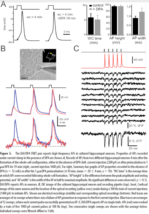
I have been itching to post about this work since David DiGregorio presented it at a meeting at Janelia last year. His group’s results, Submillisecond Optical Reporting of Membrane Potentials In Situ Using a Neuronal Trace Dye, were published in the Journal of Neuroscience last week.  Their method of optical voltage sensing is the first one that looks like its ready for “prime-time” action outside of the labs of developers of these sorts of techniques.  It has sufficient speed (<1 ms resolution), sensitivity (25% dF/F per 100mV), and limited membrane perturbation to see single action potentials, without dramatically altering the shape of these currents.  


Membrane depolarization causes DPA to rapidly partition to the inner membrane leaflet, quenching DiO.

Membrane depolarization causes DPA to rapidly partition to the inner membrane leaflet, quenching DiO.


Like previous methods, Bradley et al. use voltage-dependent membrane partitioning of dipicrylamine (DPA), a charged small molecule, to quench a fluorophore via FRET.  Previously, high-concentrations of DPA were required to have a reasonable signal change, which caused toxicity, increased membrane capacitance and slowed voltage transients.  By using DiO, a lipophilic neuronal tracer, as the fluorophore, the DPA concentration could be reduced to 1uM, while retaining sufficient optical sensitivity for action potential detection.

Picture 9


Photoactivated Transcription Revisted

14 07 2009

Looks like there has been some new results in the field of photoactivated transcription.  Unlike the fully genetically-encoded systems reviewed in a Journal Club, this uses a hybrid genetic and small molecule approach. In Doxycycline-dependent photoactivated gene expression in eukaryotic systems, Cambridge et al. add the photolabile protecting groups to doxycyclin derivatives, which then function as photoactivatable switches in the commonly used Tet-on system. Dr. Dan O’Connor described the technique as “the path of least resistance to photoactivated transcription.” 


Local (left) and global (right) GFP expression following optical uncaging of cyanodoxycyclin

Local (left) and global (right) GFP expression following optical uncaging of cyanodoxycyclin


      The authors were able to get robust gene expression with standard UV irradiation, but also were able to uncage sufficient cyanodoxycycline with two-photon illumination to cause highly localized gene expression in cultures.  In live tadpoles, they stuck to UV for the greater efficiency.

     The standard caveats of the tet system apply. The off-state still has a bit of residual gene expression, which is fine for some applications (like fluorescent tagging), and a dealbreaker for others (cre induction). Drug delivery takes time and comes with diffusion, penetration and clearance issues.  UV penetration through deep tissue is going to be a big technical hurdle to overcome to apply this to full-grown mammals. Blasting living tissue with high power UV usually isn’t a good idea. Despite these caveats, the system clearly works and I’d bet the authors are already applying the system to some next-step applications and biological questions. The potential of selectively turning on genes in functionally identified neurons via light is enormous.  It is one of the most likely eventual avenues into possible optical activation or suppression of specific thought patterns (at least if you are willing to squirt virus into your brain and eat a bunch of nasty antibiotics).


Infrared fluorescent proteins

8 05 2009

Hunting for new fluorescent proteins in the coral reefs of the Caribbean and Australia is a task that a lucky few researchers have managed to get funding for. Scuba diving in some of the world’s most beautiful places; it’s not a bad gig, if you can get it.  Most fluorescent protein scientists are confined to a lab, mutating existing fluorescent proteins from jellyfish and coral. Shifting their excitation and emission spectra has allowed multiple fluorescent proteins to be used as molecular highlighters at the same time, since their colors are distinct from each other. Some members of this palette are shown in Brain Windows top image bar.  After over a decade of research, the spectrum is pretty well covered.  Except for one area…  The infrared.

The near-infrared band is an area of enormous importance to scientific researchers, because is contains the spectral window where the body becomes transparent. Hemoglobin in the blood strongly absorbs visible wavelengths shorter than 650nm, while water absorbs wavelengths above 900nm.  If a fluorescent protein could be found, or engineered to have excitation and emission within this window, we could use it to peer much deeper into the body. The near-IR light penetrates much more easily into and out of the tissue.  This is easily seen by pressing a flashlight against your hand.  Only the deep red light passes through. The quest for an infrared fluorescent protein has preoccupied several labs for a decade.

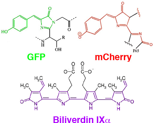
Efforts to push FPs out to the infrared resulted in mCherry, mPlummKate, among others.  The further red-shifting of these proteins is constrained by the space limitations of the beta-barrel structure of GFP-like proteins.  In general, the longer the resonance chain of the chromophore, the longer the wavelength of the chromophore’s excitation and emission spectra.  It has been difficult to extend the FP spectra beyond 650nm without adding an additional bond to the resonance structure, for which there is little space left in the protected center of the barrel.  


Wavelength of excitation and emission is longer in larger resonance structures.

Wavelength of excitation and emission is longer in larger resonance structures.

In Mammalian Expression of Infrared Fluorescent Proteins Engineered from a Bacterial Phytochrome Shu et al. of Roger Tsien’s lab, looked beyond traditionally used fluorescent proteins to extend the palette into the near-IR.  They engineered a protein, IFP, which binds biliverdin, a natural product involved in aerobic respiration (and similar in structure to the phytocyanobilins discussed in our Photoactivated Transcription journal club post). Biliverdin is non-fluorescent in solution, but when bound to IFP, it is rigidized and becomes fluorescent, with excitation at 684nm and emission at 708nm.  IFP can then be fused to the protein of interest and visualized through thick absorbent tissue.  Even the liver, which is dense with heme, is easily seen through the skin when labelled with IFP.  


IFP1.1 expressed in mouse liver is clearly visible through the skin 


IFP1.1 expressed in mouse liver is clearly visible through the skin


IFP1.4 should immediately push forward the field of in-vivo imaging of cancer and other diagnostics, at least in animal models.  It’s not clear yet how useful it will be for in-vivo brain imaging, they show cultured neurons expressing IFP1.4 become fluorescent upon biliverdin application, but can biliverdin be effectively delivered to neurons in vivo? Like channelrhodopsin, there may be sufficent amounts of endogenous co-factor to make the protein useful without exogenous application. Perhaps of greater importance is the new engineering avenues IFP opens up.  This is an entirely new, two-component scaffold, with different characteristics from GFP that protein engineers will be able to optimize and exploit.   Over the next decade, IFP may spawn as diverse a set of tools as GFP has over the previous one.


Background : Perceval, the ATP:ADP sensor

12 03 2009

Recently, Brain Windows mentioned the report A genetically encoded fluorescent reporter of ATP:ADP ratio. We invited Dr. Jim Berg, the lead author of the study to provide additional background to our readers. Below, Jim provides a fascinating look at rationale behind sensor development.  I really like that they came at this problem with a biological question in mind, something I would recommend before anyone start the development of a genetically encoded indicator.


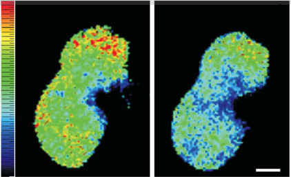
A pixel-by-pixel ratio of the 490 nm excitation image by the 430 nm excitation image from two cultured HEK293 cells expressing Perceval during control conditions (left) and after 40 min of metabolic inhibition with 5 mM 2-deoxyglucose (right)

A pixel-by-pixel ratio of the 490 nm excitation image by the 430 nm excitation image from two cultured HEK293 cells expressing Perceval during control conditions (left) and after 40 min of metabolic inhibition with 5 mM 2-deoxyglucose (right)


Here’s a little insight into why we decided to develop a fluorescent sensor for cellular energy, and how Perceval evolved. One of the primary research interests of the Yellen lab is the interaction between diet and epilepsy. The ketogenic diet, a high fat, low carbohydrate regimen, is remarkably effective at reducing seizure number. We are investigating how the transition in brain metabolism from glucose to a mixture of glucose and ketone bodies (the metabolically active byproduct of fat metabolism) could lead to a change in neuronal excitability. Previously, we described how acute application of ketone bodies reduces the excitability of substantia nigra neurons, an effect that relies on the opening of ATP-sensitive potassium (KATP) channels. Our hypothesis is that the inhibition of glycolysis by ketone body metabolism leads to a reduction in sub-membrane ATP, resulting in an opening of KATP channels and a decrease in neuronal excitability. This relies on the controversial idea that sub-membrane ATP is provided by glycolysis (possibly by glycolytic enzymes tethered to the membrane), and that the diffusion of ATP is restricted between the submembrane space and bulk cytoplasm, and concept known as “compartmentation”. To fully test this hypothesis, we required an optical sensor for ATP levels.

When planning these experiments, our first thought was to use Luciferase to detect different subcellular ATP levels. For a number of reasons, primarily Luciferase’s weak signal, we decided that a fluorescent sensor for ATP would be much more useful for our application. Our initial approach was a FRET-based design, with CFP and YFP tethered to a bacterial periplasmic binding protein that dimerized upon ATP application. Although these sensors gave some encouraging results, we never got the change in signal that would be required for cellular assays. We then adopted the ‘circularly permuted fluorescent protein (cpFP) approach that had previously produced sensors for calcium (pericam) and hydrogen peroxide (HyPer). We inserted the yellow fluorescent protein cpmVenus into the loop of the bacterial ATP binding protein, GlnK1 (involved in the regulation of ammonia transport) and found that application of small amounts of ATP to the purified sensor led to a substantial change in the excitation spectrum of the sensor. The affinity of the sensor for ATP was extremely high, orders of magnitude more sensitive than would be appropriate for cellular assays. We also found that our sensor responded to ADP application, only with a much smaller fluorescence change. It was then that we determined that these two perceived negatives (too high affinity and ADP binding) would lead to a sensor that reports the ratio of ATP to ADP. In a bit of good fortune, our design for an ATP sensor had in fact given us a sensor for the more valuable ATP:ADP ratio. After tinkering with our sensor by semirandom mutagenesis of the GlnK1 portion of the protein, we expressed the improved sensor, which we named Perceval (for permuted reporter of cellular energy value) into cultured cells and monitored a change in fluorescence with metabolic inhibition.

Right now, we are excited to use Perceval to investigate neuronal/glial metabolism in mammals. We may target subcellular ATP by either tethering Perceval to a membrane protein, or by using TIRF microscopy. In addition, we are continuing to design improved versions of Perceval, as well as sensors for other metabolic intermediates. We also hope that these sensors will be useful in applications beyond neuronal metabolism, from studies of cancer cells to bacterial metabolism.


Symposium : A Revolution in Fluorescence Imaging

11 02 2009

header-jellyfish

This coming Tuesday and Wednesday (Feb 17th & 18th) at UCSD, there will be a symposium honoring Roger Tsien, featuring presentations from 32 former and current members of the Tsien Lab. The topics are quite diverse, concentrated in genetically-encoded indicators, but also featuring fluorescent cell penetrating peptides for cancer therapy, photophore ligases for imaging synaptic development, and even a radical new design for the internal combustion engine.

The quality of speakers and subjects looks to be outstanding.  Here is a complete schedule.  You may notice that at 11:15 AM on Tuesday in Price Center East Ballroom, I will be presenting recent progress we have made in the development of genetically-encoded calcium indicators and their application to in vivo imaging.  Don’t miss that one!  🙂  Roger’s talk, which will assuredly be equal parts absorbing, humorous, and illuminating, is at 4pm Wednesday in the Price Center Theater.

If you live in Southern California and are interesting in imaging technology, there isn’t a better place to be than this symposium.  If you can’t make it, Brain Windows will have a full write-up following the event.

Here is the un-official schedule.

Tuesday February 17th – Price Center East Ballroom

9:00 -9:05 Varda  Levram -Ellisman Opening

9:05-9:15 Palmer Taylor

Designing the next generation of genetically encoded sensors

9:15-9:30 Roger Heim

FRET for compound screening at Aurora/Vertex

9:30-9:45 Amy Palmer

Designing and using genetically encoded sensors: Lessons I learned from Roger

9:45-10:00 Robert Campbell

Beyond brightness: colony screens for fluorescent protein photo stability and biosensor FRET changes

10:00-10:15 Colette Dooley

GFP sensors for reactive oxygen species: Tying up loose ends and looking forward.

10:15-10:30 Peter Wang

Fluorescent Proteins and FRET biosensors for visualizing cell motility and mechanotransduction

Fluorescent proteins in neuroscience

11:00-11:15 Brian Bacskai

Aberrant calcium homeostasis in the Alzheimer mouse brain

11:15-11:30 Andrew Hires

Watching a mouse think: Novel fluorescent genetically-encoded calcium indicators applied to in vivo brain imaging

11:30-11:45 Alice Ting

Imaging synapse development with engineered photophore ligases

11:45-12:00 Rex Kerr

3D calcium imaging in C. elegans

Clinical applications

12:00-12:15 Todd Aguilera

Activatable Cell Penetrating Peptides for use in clinical contrast agent and therapeutic development

12:15-12:30 Quyen Nguyen

Surgery with Molecular Fluorescence Imaging Guidance

Fluorescent probes (Chemistry)

1:30-1:45 Tito Gonzalez

Voltage-Sensitive FRET Probes & Applications

1:45-2:00 Paul Negulescu

From watching ions to moving them

2:00-2:15 Timothy Dore

Roger-Inspired Photochemistry: Releasing Biological Effectors with 2PE

2:00-2:15 Joe Kao

Electron Paramagnetic Resonance Imaging in Living Animals

2:15-2:30 Brent Martin

Chemical probes for studying protein acylation

2:30-2:45 Jianghong Rao

Non-GFP based probes for imaging of the hydrolytic enzyme activity

Cellular research with and without Fluorescent probes

3:15-3:30 Carsten Schultz

Cell membrane repair visualized by GFP fusion proteins

3:30-3:45 David Green

Transcriptomes and Systems Biology: application to early mammalian embryogenesis

3:45-4:00 Clotilde Randriamampita

Paradoxical aspects of T cell activation revealed with fluorescent proteins

4:15-4:30 Wen-Hong Li

Studying dynamic cell-cell communication in vivo by Trojan-LAMP

4:30-4:45 Martin Poenie

Aim and Shoot: Two roles for dynein in T cell effector function

4:45-5:00 Gregor Zlokarnik

From bla to blah, blah in 20 years

5:00-5:15                        James Sharp

President, Zeiss MicroImaging Gmbh

February 18 2009 – Leichtag 107

Cellular research with and without fluorescent proteins

9:00-9:15 David Zacharias

Fluorescent Proteins, Palmitoylation and Cancer: two out of three ain’t bad

9:15-9:30 Jin Zhang

Visualization of Cell Signaling Dynamics: A Tale of MAPK

9:30-9:45 Paul Sammak

Nuclear organization and movement in pluripotent stem cells measured by Histone GFP H2B

Branching out

9:45-10:00 Yong Yao

NIH Toolbox Program

10:00-10:15 Oded Tour

The Tour Engine – A novel Internal Combustion Engine with the potential to boost efficiency and cut emissions

Into the future

10:45-11:00 Xiaokun Shu

Visibly and infrared fluorescent proteins: photophysics and engineering

11:00-11:15 Michael Lin

Engineering fluorescent proteins for visualizing newly synthesized proteins and improving FRET-based biosensors

11:15-11:30 Jeremy Babendure

Training our next generation of Fluorescent Protein Enthusiasts

Main Event – Price Center Theater

4:00-5:00 Roger Tsien

Chancellor invitational lecture 2008 Nobel Prize in Chemistry