G-CaMP5 is finally published!

3 10 2012

The paper on G-CaMP5 has been published.

Optimization of a GCaMP Calcium Indicator for Neural Activity Imaging

Genetically encoded calcium indicators (GECIs) are powerful tools for systems neuroscience. Recent efforts in protein engineering have significantly increased the performance of GECIs. The state-of-the art single-wavelength GECI, GCaMP3, has been deployed in a number of model organisms and can reliably detect three or more action potentials in short bursts in several systems in vivo. Through protein structure determination, targeted mutagenesis, high-throughput screening, and a battery of in vitro assays, we have increased the dynamic range of GCaMP3 by severalfold, creating a family of “GCaMP5” sensors. We tested GCaMP5s in several systems: cultured neurons and astrocytes, mouse retina, and in vivo in Caenorhabditischemosensory neurons, Drosophila larval neuromuscular junction and adult antennal lobe, zebrafish retina and tectum, and mouse visual cortex. Signal-to-noise ratio was improved by at least 2- to 3-fold. In the visual cortex, two GCaMP5 variants detected twice as many visual stimulus-responsive cells as GCaMP3. By combining in vivoimaging with electrophysiology we show that GCaMP5 fluorescence provides a more reliable measure of neuronal activity than its predecessor GCaMP3. GCaMP5 allows more sensitive detection of neural activity in vivo and may find widespread applications for cellular imaging in general.

Image

This is the best fully-characterized GECI available, but publication of the paper was repeatedly delayed. Why? Because reviewers viewed it as ‘too incremental’ of an upgrade, and not worth publishing in a prominent journal (no, I’m not talking Nature or Science level) when the plasmids are already available.

A friendly suggestion for authors and future GCaMP6+ reviewers : You can’t have it both ways. If you want access to the best molecular tools before publication (GCaMP5 has been available for over a YEAR), you cannot turn around and say its not worth publishing because you already have the plasmid. Multiple post-docs spent years of their lives developing and carefully testing this tool. They deserve a quality publication for their efforts. Furthermore, the rigorous performance data collected NEEDS to be available to current and future users. Finally, there is no doubt that this will be a highly cited and viewed paper in whatever journal were to publish it. Our GCaMP3 paper already has 182 citations in less than 3 years, this may do even better.


Three Cheers for GCaMP : Optogenetic Brain Reading

9 11 2009

Three papers are out online in Nature Methods that show big improvements in calcium imaging with genetically encoded sensors.  They are are based on the fluorescence intensity indicator, GCaMP.   GCaMP, first developed by Junichi Nakai, consists of a GFP that has been circularly permuted so that the N and C termini are fused and new termini are made in the middle of the protein.  Fused to one terminus is calmodulin and the other is a peptide, M13, that calmodulin (CaM) binds to in the presence of calcium. The name is supposed to look like GFP with a CaM inserted into it, G-CaM-P.  Normally the GFP is dim, as there is a hole from the outside of its barrel into the chromophore.  Upon binding calcium, this hole is plugged and fluorescence increases.

Crystal structure of GCaMP2

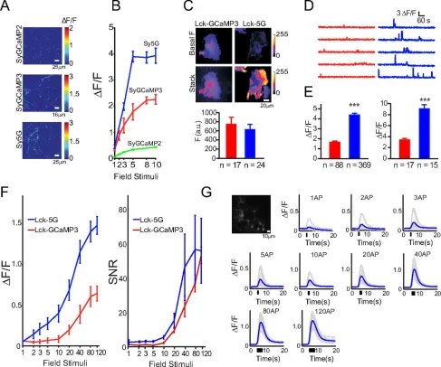
The first paper, A genetically encoded reporter of synaptic activity in vivo, from Leon Lagnado’s group, targets GCaMP2 to the outer surface of synaptic vesicles. This localization allows the fluorescence signal to be confined to the presynaptic terminal, where calcium fluxes in response to action potentials are high.  This targeting improves the response magnitude of GCaMP2 and permits the optical recording of synaptic inputs into whatever region of the brain one looks at.  They demonstrate the technique in live zebrafish.

In the second paper, Optical interrogation of neural circuits in Caenorhabditis elegans, from Sharad Ramanathan’s group, GCaMP2 has been combined with Channelrhodopsin-2 to perform functional circuit mapping in the worm.   Since the worm’s structural wiring diagram has been essentially solved, functional data could say much about how “thick” the wires between each cell are.  Unfortunately, with GCaMP2, the responses are too slow and weak to distinguish direct from indirect connections.

Finally, we have published a paper, Imaging neural activity in worms, flies and mice with improved GCaMP calcium indicators, describing the improved GCaMP3.  This indicator has between 2-10x better signal to noise than GCaMP2, D3cpv and TN-XXL, depending on the system you are using.  It’s kinetics are faster and it is more photostable than FRET indicators, and the responses are huge.  When expressed in motor cortex of the mouse, neuronal activity is easily seen directly in the raw data.  Furthermore, the sensor can be expressed stably for months, making it a potential tool for observing how learning reshapes the patterns of activity in the cortex.

Screen shot 2009-11-09 at 7.19.27 PM

Imaging of mouse motor cortex (M1) expressing the genetically-encoded calcium indicator GCaMP3 through a cortical window. After 72 days of GCaMP3 expression, large fluorescence transients can be seen in many neurons that are highly correlated with mouse running.

GCaMP3 is not perfect. It cannot reliably detect single action potential in vivo in mammals, though I doubt that any existing GECI can. Work continues on future generations of GCaMP that may achieve 100% fidelity in optical reading of the bits in the brain. However, there is considerable evidence from a number of groups that have been beta-testing the sensor, including the Tank lab of “quake mouse” fame, that it is a significant leap forward and unlocks much of the fantastic and fantasized potential of genetically-encoded calcium indicators.

Screen shot 2009-11-09 at 7.20.12 PM

Comparison of fluorescence changes in response to trains of action potentials in acute cortical slices.

I will try to post a more complete writeup of GCaMP3 for Brain Windows soon, with an unbiased eye to its strengths and weaknesses.  We worked very hard to carefully characterize this sensor’s effects on cellular and circuit properties.  If you have any questions about GCaMP3, please post them to the comments.

For further info about strategies for GECI use and optimization, check out our previous paper, Reporting neural activity with genetically encoded calcium indicators in Brain Cell Biology.

The official press release from HHMI regarding GCaMP3 is available here.